Sunday, May 16, 2010

Episcleritis


Pathophysiology

The pathophysiology is poorly understood. The inflammatory response is localized to the superficial episcleral vascular network, and histopathology shows nongranulomatous inflammation with vascular dilatation and perivascular infiltration.

The 2 clinical types are simple and nodular.

The most common type is simple episcleritis, in which there are intermittent bouts of moderate-to-severe inflammation that often recur at 1- to 3-month intervals. The episodes usually last 7-10 days, and most resolve after 2-3 weeks. Prolonged episodes may be more common in patients with associated systemic conditions. Some patients note that episodes are more common in the spring or fall. The precipitating factor is rarely found, but attacks have been associated with stress and hormonal changes.

Patients with nodular episcleritis have prolonged attacks of inflammation that are typically more painful than simple episcleritis. Many patients with nodular episcleritis have an associated systemic disease.

Dacryocystitis


The naso-optic fissure is the source of origin of the lacrimal drainage system. The ectoderm in this region thickens and becomes embedded in the mesenchyme between the lateral nasal and maxillary processes. This cord of ectoderm subsequently canalizes and opens into the conjunctival fornix prior to opening into the nasal vestibule. Frequently, this opening into the nasal cavity is incomplete at birth. Canalization of the lacrimal excretory system begins in the superior portion first and is segmental, only later coalescing to form a continuous lumen. The canaliculi, which develop as outpouchings from the solid cord of ectodermal tissue prior to canalization, also canalize prior to the vertical portions of the nasolacrimal duct.

Many variations in the anatomy of the lacrimal drainage system have been noted. Normally, tears drain into the lacrimal system through two puncta, one present in the upper lid and the other in the lower lid. More commonly, the lower punctum lies slightly temporal to the upper punctum.

The connections from the puncta to the lacrimal sac are called canaliculi. These canaliculi have a short vertical segment, averaging 2 mm in length, and a longer horizontal segment, averaging 10-12 mm in length.

An ampulla connects the vertical and horizontal segments. The individual canalicular horizontal segments join to form a common canaliculus in 90% of patients. This common canaliculus dilates, forming the sinus of Maier just lateral to the lacrimal sac.

A fold of mucosa known as the valve of Rosenmüller marks the junction of the lacrimal sac and the common canaliculus. The lacrimal sac lies in the bony lacrimal fossa derived from the lacrimal and maxillary bones. The average width of the sac is approximately 6-7 mm and the length varies from 12-15 mm. The mucosa of the sac is lined by pseudostratified columnar epithelium with substantial amounts of lymphoid and elastic tissue interposed within the connective tissue layer. The sac is normally irregular and flat in shape with a collapsed lumen.

The lacrimal sac is covered on its outer surface by the lacrimal fascia of the periorbita. This fascia splits to envelop the lacrimal sac between the attachments of the lacrimal fascia to the anterior and posterior lacrimal crests. The lacrimal sac mucosa only loosely adheres to the lacrimal fascia. However, posterior to the sac are the deep heads of the pretarsal and preseptal orbicularis muscles. Anteriorly, the medial canthal tendon covers the upper two fifths of the lacrimal sac.

The nasolacrimal duct averages 18 mm in length and 4.5-5 mm in diameter. Multiple valves are present in the nasolacrimal duct, representing analog from the segmental canalization of the ectodermal cord that develops into the nasolacrimal duct. Of these, the most prominent valves are the valve of Taillefer, the valve of Krause, and the valve of Hasner (located at the junction of the duct with the nasal mucosa). Like the lacrimal sac, the nasolacrimal duct is lined by pseudostratified columnar epithelium.

The lacrimal, maxillary, and ethmoid bones form the bony nasolacrimal canal. The bulk of the duct is contributed by the maxilla, anteriorly, laterally, and posteriorly. The lacrimal bone forms the medial wall superiorly, and the inferior concha of the ethmoid bone forms the medial wall of the canal inferiorly. The mucosal opening of the nasolacrimal duct under the inferior turbinate lies 5-8 mm from the anterior tip of the inferior turbinate. The lacrimal bone and the nasal process of the maxilla make up the lacrimal fossa equally. The anterior and posterior lacrimal crests form the anterior and posterior borders of the lacrimal fossa, respectively.

The dimensions of the lacrimal fossa are 4-8 mm in width, 15 mm in height, and 2 mm in depth. Ethmoid air cells in approximately 40-60% of patients separate the lacrimal fossa from the nasal cavity, although considerable variability exists in the number and location of these air cells. The lacrimal sac fossa lies at the level of the anterior tip of the middle turbinate.

Dacryo Adenitis


The pathophysiology is not understood completely. Yet, infectious dacryoadenitis is thought to be caused by ascension of an inciting agent from the conjunctiva through the lacrimal ductules into the lacrimal gland.

Corneal Abrasions

 
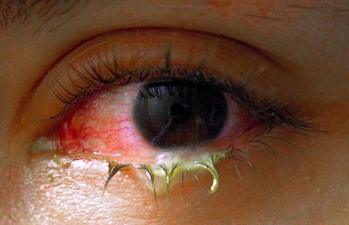
Abrasion is a defect in the surface of the cornea that is limited to the epithelial layers and that does not penetrate the Bowman membrane. In some cases, the bulbar conjunctiva is also involved. Corneal abrasion results from physical or chemical trauma. Severe corneal injuries can also involve the deeper, thicker stromal layer; in this situation, the term corneal ulcer may be used.

The conjunctival response to corneal wounding has been known since Mann first observed that peripheral corneal abrasions heal by the sliding of limbal cells to cover the epithelial defect.1 This response is split into 2 phases: (1) the response of the limbal epithelium, which is the source of the corneal epithelial stem cells, and (2) the response of the conjunctival epithelium itself.

Under normal circumstances, the limbal epithelium acts as a barrier and exerts an inhibitory growth pressure that prevents the migration of conjunctival epithelial cells onto the cornea. Like the rest of the surface of the body, the conjunctiva and the cornea are in a constant state of turnover. Corneal epithelial cells are continuously shed into the tear pool, and they are simultaneously replenished by cells moving centrally from the limbus and anteriorly from the basal layer of the epithelium. Movement from the basal to superficial layers is relatively rapid, requiring 7-10 days; however, movement from the limbus to the center of the cornea is slow and may require months.

This normal physiologic process is exaggerated in the case of a corneal abrasion. During corneal healing of a lesion, corneal epithelial cells become flattened, they spread, and they move across the defect until they cover it completely. Cell proliferation, which is independent of cell migration, begins approximately 24 hours after injury. Stem cells from the limbus also respond by proliferating to give rise to daughter cells called transient amplifying cells. These cells migrate to heal the corneal defect and proliferate to replenish the wounded area.

The observation of limbal pigment migrating onto the clear cornea provided additional evidence of this process. The concept that the limbal cells form a barrier to conjunctival cells was supported further by the observation that rabbit eyes treated for 120 seconds with N -heptanal, which removed the corneal and conjunctival epithelium but left the limbal basal cells intact, healed with the corneal epithelium and had unvascularized corneas. However, when the entire limbal zone was surgically removed along with N -heptanal treatment, corneal vascularization and conjunctivalization was observed.

Demonstration of the centripetal migration of limbal cells (marked by India ink) provided more direct evidence of this concept. These cells migrate in masses as a continuous, coherent sheet, with most cells retaining their positions relative to each other, much like the movement of a herd of cattle.

Rearrangement of intracellular actin filaments plays a role in movement. Cell migration can be inhibited by blocking polymerization of actin, indicating that actin filaments actively participate in the mechanism of cell motion. Some authors believe that conjunctival and limbal epithelial cells may contribute to the regeneration of corneal epithelium. Marked proliferative responses in the conjunctiva after a central corneal epithelium abrasion have been described.

Why the conjunctival epithelium should proliferate in response to a central corneal wound is unknown. One possibility is that the proliferation replenishes the number of goblet cells, which decreases by up to 50% after corneal wounding. However, proliferation occurs at high levels in the bulbar conjunctiva, which contains few if any goblet cells. The apparent decrease in cell number is more likely the result of mucin secretion rather than actual loss of goblet cells. Alternately, conjunctival cells may migrate into the limbus or cornea to help replenish the wound area. No firm data suggest that conjunctival epithelium migrates onto the corneal surface in the presence of intact limbal epithelium. Last, healing of the corneal epithelial wound is not complete until the newly regenerated epithelium has firmly anchored itself to the underlying connective tissue.

Permanent anchoring units are not formed until the wound defect is covered completely. Epithelial cells migrate rapidly and develop strong, permanent adhesions within 1 week when the basement membrane is regularly formed and released during the cell migration process.

Although transient attachments are regularly formed and released during the cell migration process, formation of normal adhesions takes 6 weeks, according to Dua et al.2 Tiny buds of corneal epithelium are present along the contact line between the normal corneal epithelium and the migrating conjunctival epithelium. These buds arise from the corneal epithelium, and normal corneal epithelium appears to replace the conjunctival epithelium by gradually pushing it toward the limbus. The magnitude and extent of both the conjunctival and corneal regenerative responses to a corneal abrasion are correlated with the size of the wound. Large erosions were reported to induce a pronounced response in the rate of epithelial cell migration and mitosis at the limbus.

Insults caused by chemical injuries, Stevens-Johnson syndrome, contact lens–induced keratopathy, and aniridia result in limbal damage. These insults cause delayed healing of the cornea, recurrent epithelial erosions, corneal vascularizations, and conjunctival epithelial ingrowth.

Role of the epithelial defect

A long-standing clinical observation is that corneal abrasions and bacterial corneal infections do not occur in patients with an intact, healthy epithelium. Bacterial keratitis and abrasions develop in 1 of 3 types of patients: (1) those with trauma to the cornea; (2) those with epithelial defects due to intrinsic disease (eg, dry eye, exposure keratitis, neurotrophic keratitis, postinfectious persistent epithelial defects); and (3) those who wear contact lenses, especially extended-wear hydrophilic lenses.

The common feature among the 3 groups is a defect in the corneal epithelium to which the bacteria must adhere to start the infection. Mechanisms underlying the development of epithelial defects in the first 2 groups are self-evident. In the third group, contact lenses may lead to epithelial injury in different ways. The cornea can be injured by insertion or removal of the lens, by trauma from defects in or deposits on the lens, by lens-induced hypoxia, or by chemical toxicity from contact-lens disinfectants.

Defects in the epithelium need not be full thickness. Overnight wearing of soft lenses, which do provide inadequate oxygen transmissibility to prevent hypoxia, causes superficial desquamation of epithelium and increases the propensity for abrasions. Corneal swelling induced by overnight wearing of contact lenses is the most important factor. The cornea normally swells 2-4% during sleep. With a contact lens, overnight swelling is increased to an average of 15%, and gross stromal edema can be present on awakening. In some patients, induced corneal swelling can be sufficient to cause bullae; these can rupture, leading to epithelial defects.

Chalazion

Click to view full image- chalazion

A chalazion is a lump of the lid that is caused by obstruction of an oil gland within the upper or lower eyelid. This lump may increase in size over days to weeks and may occasionally become red, warm, or painful.

The gland involved in the formation of a chalazion is a modified oil gland that lies within the eyelid. There are about 40 of these glands within each of the upper and lower lids. These glands secrete oil into the tears. When one of these glands becomes blocked, it can increase in size and cause a visible lump.

Although a sty is also a lump in the eyelid caused by obstruction of an oil gland, a chalazion is not a sty. A sty, or hordeolum, represents an acute infection of the gland. A chalazion is not an infection but is an inflammation of the area. Inflammation is a process in which the body reacts to a condition and produces a swelling, redness, pain, or warmth. A sty is usually more painful than a chalazion and may look infected.

Bacterial Conjunctivitis

Click to view full screen- bacterial conjunctivitis

Conjunctivitis (also called pink eye or madras eye refers to inflammation of the conjunctiva (the outermost layer of the eye and the inner surface of the eyelids).[1] It is most commonly due to an infection (usually viral, but sometimes bacterial or an allergic reaction.

Arcus senilis

click to view full screen arcus senilis  
Arcus senilis (or arcus senilis corneae) is a white or gray opaque ring in the corneal margin (peripheral corneal opacity), white ring around the iris. It is present at birth but then fades; however it is quite commonly present in the elderly. It can also appear earlier in life as a result of hypercholesterolaemia.

Angular Blepheritis

click to vewi full screen- blepharitis

(pronounced /blɛfərˈaɪtɨs/ BLEF-ər-EYE-tis) is an ocular condition characterized by chronic inflammation of the eyelid, the severity and time course of which can vary. It can onset acutely resolving without treatment within 2–4 weeks (this can be greatly reduced with lid hygiene) but more generally is a long standing inflammation varying in severity. It may be classified as seborrhoeic, staphylocccal, mixed, posterior or meiobomitis, or parasitic.

click to veiw full screen - internal hordeolum

Alkali Burn 3


Damage to tissue caused by exposure to an alkaline compound such as lye. Treatment includes washing the area with copious amounts of water to remove the chemical. It takes more water to irrigate an alkali burn than an acid burn. The victim is immediately taken to a medical facility for assessment of tissue damage. 

Alkali Burn 2


Damage to tissue caused by exposure to an alkaline compound such as lye. Treatment includes washing the area with copious amounts of water to remove the chemical. It takes more water to irrigate an alkali burn than an acid burn. The victim is immediately taken to a medical facility for assessment of tissue damage. 

Alkali Burn (EYE)

Disease images, eye diseases, alkali burn

Saturday, May 15, 2010